Monday, February 2, 2009

Microbiology experiments

During the past 16 weeks, we have done a series of experiments involving microbiology. The following is to help you remember the experiments which we’ve done in microbiology. (As such, the procedures are all in active voice)
Cell culture
Learning how to subculture a continuous cell line is integral in any field of microbiology. This experiment basically teaches us how to subculture a continuous cell line, what the functions of solutions such as trypsin, Phosphate buffered saline, and growth mediums are etc. Also, we will see what a confluent monolayer should look like.
Functions of solutions
Phosphate buffered saline (PBS)-> Due to the fact that PBS is isotonic and non-toxic to cells, it is usually used to rinse containers containing cells, dilute substances etc.
Trypsin-> Used to re-suspend cells which have adhered to the cell culture’s dish wall by lysing the bonds between the proteins and the cell culture’s flask wall
Growth medium-> Contains peptide hormones or hormone-like growth factors which promote healthy cell growth; also neutralizes proteases such as trypsin
Summary of procedures
1. Decant medium from cell flask
2. Wash with PBS
3. Add trypsin
4. Incubate the cells in the flask till cells detach
5. Aspirate cell suspension to disperse cell aggregates
6. Transfer some of the suspension into a new cell flask
7. Incubate the flask with cells


Top: A confluent monolayer
Http://www.devicelink.com/mddi/archive/98/04/013.html
Cell counting
Cell counting is also an invaluable tool in microbiology. It is in cell counting that we derive the concentration of cells, in cells/ml from. Using the formula,
Cells/ml = Average cell count per square x dilution factor x 104
Summary of procedures
1. For every 5ml of cells, add 45ml of trypan blue. Mix them and leave them to stand
2. Transfer a small amount of solution onto the haemocytometer
Basically, just count all the cells in the big square in the middle, along with the 4 big corner squares and average them to get your “average cell count per square” value.


http://en.wikipedia.org/wiki/File:Haemocytometer_Grid.png
Top: A haemocytometer chamber.
Red square = 1.0000 mm2
Green square = 0.0625 mm2
Yellow square = 0.040 mm2
Blue square = 0.0025 mm2
All squares have a depth of 0.1 mm if a cover slip is in place
Each big (red) square has a volume of 0.1mm3, or 0.0001cm3. As we want the concentration to be in cm3 (ml), we multiply our value by 10,000 as 0.0001 x 10,000 = 1

Viral infection and amplification
We can culture viruses by getting them to infect host cells. By infecting the host cells with virus, we would be able to observe what the viruses do to the infected hosts. These changes are called cytopathic effects (CPE). As CPE varies from host to host, and from virus to virus, we can use that to determine the species of the virus.
After we get a culture of virus, we should then amplify our virus pool so that we can conduct further research.


Summary of procedures
Viral infection
1. Decant medium from flask and wash monolayer with virus diluent
2. Decant the virus diluent
3. Clean and disinfect the surface of the cryovial with 70% alcohol
4. Transfer the virus from the cryovial to the virus suspension
5. Incubate the virus suspension
6. After 1 hour, remove excess inoculum and rinse monolayer with virus diluent
7. Add maintenance medium
8. Incubate
Viral amplification
9. Decant medium from big flask, then wash with virus diluent
10. Transfer the medium from the original infected flask to the new, big flask
11. After 1 hour, remove the excess inoculum and rinse with virus diluent
12. Add maintenance medium


http://library.thinkquest.org/10607/media/cell-virus.gif
Top: Viral infection
Plaque assay
Since viruses are too numerous to be quantified by direct counting, scientists have come up with plaque assay, which is an indirect method to count viruses. The formula for virus concentration, in plaque forming units (pfu)/ml, is given as,
Virus concentration = (number of plaques) x (dilution factor(s)) pfu/ml
But before we can do the plaque assay, we must first make serial dilutions of a given virus sample.
Also, there is a new solution:
Carboxymethyl cellulose(CMC) solution-> Used to increase the viscosity of the solution
Summary of procedures
Serial dilution
1. Take 10 tubes and label them from 1 to 10. Then add virus diluents into each tube.
2. Transfer x ml of the virus suspension into the tube labeled 1.
3. Transfer the same amount of virus suspension in tube 1 into 2. Repeat till tube 10
Plaque assay
1. Label 2 wells 6, 2 wells 7 and so on until 10. Label the last 2 as controls
2. Decant the medium from the trays of cells and wash them with virus diluent
3. Decant the virus diluent
4. To the well labeled 6, transfer x ml of virus suspension from tube 6. Add the same amount for the other wells. Ex: If you add 0.5ml of virus suspension from tube 6 to well 6, add 0.5ml of virus suspension from tube 7 to well 7
5. Incubate the wells
6. After 1 hour, remove the excess inoculums and rinse the monolayer with virus diluent
7. The maintenance medium and CMC solutions together. Add the same amount of the mixture to each well.


http://upload.wikimedia.org/wikipedia/commons/f/fb/Plaque_assay_dilution_series.jpg
Top: 4-fold dilution series from top-left to bottom-right, stained with crystal violet. Living cells are not stained whereas dead cells are.

ELISA

An ELISA is used to detect the presence of antibodies or antigens in a sample. In our experiment, we used ELISA to test for the presence of a specific antigen. To test for a presence of a specific antigen, we attach its complementary antibody onto the walls of the wells. (This means that we attach an antibody which will “absorb” the antigen onto the walls of the wells)

Please click here to learn about the basics of ELISA.

Summary of procedures
Antigen dilution
1. Add the sample diluted to their correct wells
2. Incubate
Wash procedure
3. After 30 minutes, aspirate the contents of the wells
4. Wash with buffer, let it stand for 1 minute, and decant. Repeat thrice


Add secondary antibody solution
5. Add secondary antibody
6. Incubate for 30 minutes
React substrate
7. Add substrate into well
8. Incubate in dark place for 10 minutes
9. Add appropriate stop solution and shake well
10. Read plate with plate reader

Baltimore

The baltimore classification is a virus classification system which groups viruses into families depending on their types of genome ( DNA, RNA , single-stranded , double stranded ) and the ways the viruses replicates.

Class 1 : Double stranded DNA viruses

Double stranded DNA viruses usually enter the host nucleus before it is able to replicate.These Viruses require host cell polymerases to replicate the viral genome and hence are highly dependent on the cell cycle.
Proper infection and production of progent requires that the cell be in a replication which is when the cell's polymerases is active thus the virus can then undergo cell division leading to tranformation of the cell and finally cancer. Example : poxvirus.

Class II : single stranded DNA viruses

These viruses replicate within the nucleus and form a double stranded DNA intermediate during replication.
This class of viruses are not very well studied.

Class III: Double stranded RNA viruses

This viruses replicates in the cytoplasm and does not require the hot replication polymerases as much as DNA viruses. Replication is monocistronic and includes individual , segmented genomes which means that each of the genes code for only one protein which is unlike other viruses which exhibit more complex translation.

Class IV and V : single stranded RNA viruses

There are two types of these virus, but they share the fact that replication is primarily in the cyotplasm, and that replication is not as dependent on the cell cycle as other DNA viruses.

Class IV: Single stranded RNA viruses - Positive (+) sense

Positive sense RNA viruses can be directly accessed by host polymerases to immediately form proteins. which are divided into 2 groups both reproduced in the cytoplasm.
(1)
Viruses with polycistronic mRNA where the genome RNA and is translated into a polyprotein product that is eventually cleaved to form the mature proteins.
(2)
Viruses with complex transcription , subgenomic mRNAs . ribosomal frameshifting and proteolytic processing of polyproteins might be used.and proteins produced from the same strand of RNA.


Class V: Single stranded RNA viruses - Negative (-) sense

these viruses cannot directly be accessed by host polymerases to immediately form proteins unlike positive. these viruses must be transcripted by viral polymerases into a form which is positive sense reciprocal. there are 2 groups.
(1)
contains non segmented genomes where first step in replication is transcription from negative stranded genome by the viral RNA-dependent RNA polymerase to yield monocistronic mRNAs which code for various viral proteins.replication occurs within the cytoplasm.
(2)
Contains segmented genomes which replication occurs in the nucleus and where the viral RNA-dependent RNA polymerase produces monocistronic mRNAs from each genome segment. the difference of both is the location os replication.

Class VI: positive sense single stranded RNA viruses that replicate through a DNA intermediate

Feature of this virus is the use of reverse transcriptase to convert the positive sense RNA into DNA. these viruses use DNA to create the templates of proteins instead of the RNA, which is spliced into the host genome using integrase,replication can then start with the aid of the host cell's polymerases. example : HIV

Class VII: Double stranded DNA viruses that replicate though a single stranded RNA intermediate

these viruses which are a small group have a double-stranded ,gapped genome that is subsequently filled in to form a covalently closed circle that serves as a template for production of viral mRNAs and a subgenomic RNA. the pregenome RNA servres as template for the viral reverse transcriptase and for production of the DNA genome.

Culturing viruses in the laboratory

Now that you know what is cell culture and the importance of it, this section will be about culturing viruses.

Viruses must be cultured in order to conduct research and develop vaccines and treatments, but because viruses cannot metabolize or replicate by themselves, they cannot be grown in standard microbiological broths or on agar plates thus they have be to cultured inside suitbale host cells , a requirement that complicates the detection, identification and characterization of viruses.
Virologist have developed 3 types of media for culturing viruses.

First , culturing viruses in bacteria.

Most of our knowledge of viral replication has come from research on bacteriophages.
which is quite easy to culture because bacteria are easily grown and maintained. Phages can be grown in bacteria maintained in either liquid or on agar plates.
Bacteria and phages are mixed with warm nutirents agar and poured in a thin layer across the surface of an agar plate. during incubation , bacteria infected by phages lyse and release new phages that infects nearby bacteria while uninfected bacteria grow and reproduce normally.
After incubation,the appearance of the plate includes a uniform bacterial lawn interrupted by clear zones called plagues. Which are areas where pahges have lysed the bacteria. Such plates enable the estimation of phage numbers via a technique called plaque assay in which each plaque corresponds to a single phage in the original bacterium or virus mixture.

second , culturing viruses in plants and animals.

most experiments designed to study diseases processes and immune responses.Frist discovery and isolation of virus was the discovery of tobacco mosaic vius in tobacco plants.Viruses that infect only humans raises additional ehtical complications.therefore scientists have developed alternative ways of culturing animal and human viruses using fertilized chicken eggs or cell cultures.


Third, cultuing viruses in embryonated chicken eggs

Chicken eggs are useful cutlure medium for viruses because they are inexpensive and among the largest of cells free of contaminating microbes.the yolk is nourishing which is suitbale for viruses to grow in.
So viruses are injected ionto embryonated eggs at the sites that are best suited for the virus's maintenance and replication.
Vaccines against some viruses can be also prepared in egg cultures.

Sunday, February 1, 2009

Families of viruses

Hepadnaviridae...Herpesviridae...Orthomyxoviridae...Picornaviridae...Flaviviridae

Hepadnaviridae


All of the known hepadnaviruses are hepatotropic, eg. Hepatitis B virus (HBV). infecting liver cells, and all can cause hepatitis in known host. Hepatitis inflammation of the liver. It can be caused by hepatitis viruses, other viruses and non-infectious agents such as alcohol.


Diseases Caused

1) Genus Orthohepadnavirus

Disease Natural Host
Hepatitis B (HBV) Humans, Chimps, Gibbons, Monkeys
Hepatitis B Ground Squirrels, Woodchucks, Chipmunks
Hepatitis B Woodchucks
2) Genus Avihepadnavirus

Disease Natural Host

Duck Hepatitis B Ducks, Geese
Genome
- partial ds DNA, circular, monopartite, smallest at 3.2kb in length
- two uneven strands of DNA. One has a negative-sense orientation, and the other, shorter, strand has a positive-sense orientation
- it is a group 7 virus, replication involves and RNA intermediate

Morphology

- roughly spherical, icosahedral nucleocapsid.

- Replication: Violates Central Dogma-DNA->RNA->DNA which they transcribe back into cDNA using reverse transcriptase

Clinical Symptoms

- Jaundice, fatigue, abdominal pain, nausea
*Note: This are acute symptoms.

Transmission

- Blood-borne, sexual

Prevention

- Recombinant HBV surface antigen vaccine or HBV immunoglobulin post-exposure prophylaxis

- Avoid sexual contact/ bloog contact
- Vaccines against hepatitis B were introduced in the early 80s and babies were all vaccinated.

Symptoms of Acute Infection

Symptoms usually activate 6 weeks to 6 months afterwards acknowledgment to the virus. In adults, aboriginal affection can cover abhorrence, loss of appetite, vomiting, fatigue, and belly cramps and jaundice. Persons can accept all or alone a few of the aloft signs and symptoms. 50% of adults with a new infection accept no affection. Over 90% of infants, 50% of children, and 5% of adults with astute hepatitis B will advance abiding or abiding infection. Abiding hepatitis B may advance to alarmist ache including cirrhosis and alarmist cancer.

Symptoms of Chronic Infection

Most patients do not undergo any symptoms. Abiding hepatitis B may advance to alarmist ache including cirrhosis and liver cancer.


Herpesviridae

A heterogeneous family of morphologically similar viruses, all of which contain double-stranded DNA and infect humans and a wide variety of other vertebrates as well as some invertebrates, such as oysters.Human herpesvirus infections are endemic and sexual contact is a significant method of transmission for several including both herpes simplex virus 1 and 2 (HSV-1, HSV-2), also human cytomegalovirus (HHV-5) and likely Karposi's sarcoma herpesvirus (HHV-8).


Virions are enveloped, ether sensitive, and vary up to 200 nm in diameter; the nucleocapsids are 100 nm in diameter and of icosahedral symmetry, with 162 capsomeres.The family is subdivided into three subfamilies Alphaherpesvirinae, Betaherpesvirinae, and Gammaherpesvirinae.


Symptoms of Herpes Simplex

An infection by a herpes simplex virusis marked by watery blisters in the skin or mucous membranes of the mouth, lips or genitals.

Symptoms of Varicella Zoster
An infection of Varicella zoster virus will cause fever, lesions. Lesions heal with a scab.


picture extracted from : http://health.allrefer.com/health/chicken-pox-treatment-chicken-pox.html

Orthomyxoviridae

Influenza virus is transmitted from person to person in droplets released by sneezing and coughing. Some of the inhaled virus lands in the lower respiratory tract. The cough may be persistent but the most prominent symptoms of influenza are systemic: fever, muscle aches.

Vaccines: Isolated HA gives good serological protection. Vaccines are produced by reassortment of egg-adapted strains with the required HA type. Large amounts of virus are then grown in embryonated eggs, being purified and formalin inactivated. The amount of doses given to a patient depends on the age of patient.
Orthomyxoviridae
For Orthomyxoviridae, there are 2 Influenza virus types A and B. They causes of acute respiratory illnesses. Both virus types cause epidemics of considerable morbidity and mortality.

Influenza virus A causes of all flu pandemics and infect humans, other mammals and birds
Influenza virus B infect humans and seals
Influenza virus C infects humans and pigs.


Picornaviridae


It is the oldest viruses which found in ancient Egypt at 1400 B.C. Picornaviruses are also among the most diverse viruses, with over 200 serotypes causing infections such as Polio, Hepatitis A, and the common cold. Foot-and-Mouth Disease Virus (genus: Apthovirus), which causes infections in livestock, was one of the first viruses to be recognized; it was discovered by Loeffler and Frosch in 1898.
Picornaviruses contain positive sense, single-stranded RNA that is approximately 7-8 kilobases long.
The viral RNA is infectious and replication takes place in the cytoplasm.
The virus has an IRES (Internal Ribosomal Entry Site) which distinguishes it from many other RNA viruses.
The virus is naked with an icosahedral capsid.
The capsid is one of the smallest of all viruses with a diameter of only 27-30nm.
Translation and cleavage of viral polypeptides produces eleven distinct proteins.
Flaviviridae
Symptoms of Dengue Fever & Dengue Hemorrhagic FeverDengue fever usually starts suddenly with:
- high fever
- severe headache
- vomiting
- pain behind the eyes
- muscle and joint pain
A rash appears 3 to 4 days after the start of the fever. The illness can last up to 10 days, but complete recovery can take as long as a month. Most dengue infections result in relatively mild illness, but some can progress to dengue hemorrhagic fever.
With dengue hemorrhagic fever, the blood vessels start to leak and cause:
- bleeding from the nose, mouth, and gums
- Bruising (a sign of bleeding inside the body)
In severity, the blood vessels can collapse, causing shock. Dengue hemorrhagic fever is fatal in about 5 percent of cases.

Thursday, January 29, 2009

Virus-host interactions

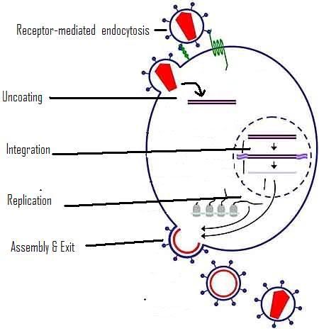
In order for a virus to infect a host, like you and I, several processes must occur involving the virus and its target cells. They are, in the following order:

1. Attachment & Entry
2. Uncoating
3. Integration
4. Replication
5. Assembly
6. Exit

Attachment & Entry

This is when a virus binds to the surface of a cell.

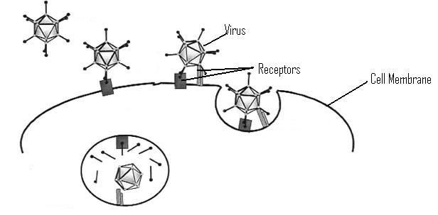
In one type of attachment, called receptor-mediated endocytosis, the virus must bind to a specific protein on the surface of the cell’s membrane, using one of its own surface proteins. Once this occurs, the cell is stimulated to take in the virus by infolding its membrane until a vesicle containing the virus is pinched off inside the cell itself.

Photobucket

Normally, this process is used to take in certain substrates that the cell uses to grow and perform its function. However, since the virus is able to replicate the signals of these substrates, the cell takes the virus in anyway.

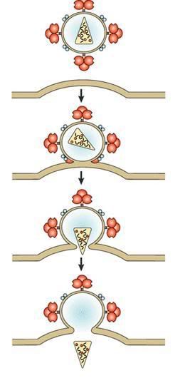
The other type of attachment, fusion, occurs without the need for the cell to perform endocytosis. However, this method requires the virus to be an enveloped virus.

When the envelope of the virus makes contact with the cell surface membrane, the two begin to fuse together. This is possible because of the fluid nature of the phospholipid bilayer(what membranes are made of). Once the fusion occurs, the virus is released into the cytoplasm of the cell.

Photobucket

Uncoating

Most viruses have a protein coat surrounding their genetic material, called the capsid. Once inside the cytoplasm of the host cell, the capsid must be broken down in order to release the genetic material and its other contents.
This is done by the host cell’s own hydrolytic enzymes. The capsid is digested, releasing the genetic material.


Integration

Integration does not happen for all types of viruses. Only DNA viruses need to have their genetic material integrated into the genome of the host cell. This is done using enzymes called restriction enzymes and integrases.

The restriction enzymes cut a section out of the cell’s genome, and the viral genome is placed in the gap using the integrases. This effectively makes the viral genome part of the cell’s genome.

Photobucket

Replication

This step is when the viral genomes are expressed to form viral proteins. DNA viruses will have their DNA transcripted and translated in the same way that the host genome produces proteins. RNA viruses can act as messenger RNA, bypassing the need for transcription.

The genome of the virus is also replicated, using the host cell’s machinery.

Assembly

The viral proteins and nucleic acids that are produced will then be transported together, and then assembled into new viruses. A single virus infecting a cell can produce many more viruses.

Exit

The exit of the new viruses can occur in two main ways.

The first method is known as budding. The newly assembled viruses move to the edge of the cell, and the viruses move out of the cell, taking a portion of the cell’s membrane with them. The stolen bit of cell membrane becomes the envelope of the virus, and will be used to aid infection of other similar cells in the future.
The other method involves cell lysis. One way this can happen is that the cell produces such a large number of viruses that the cell cannot contain them, and simply bursts, releasing the viruses. Or, the viral genome can cause the cell to destroy itself with its own enzymes, also releasing the virus.

When the viruses are released out of the cell, they move on to infect new cells, repeating the cycle.

Here is an overview of the viral lifecycle:

Photobucket

Friday, January 2, 2009

Poxviridae

The poxviridae family of viruses is the largest and most complex of viruses that have been discovered to date.

Poxviridae are so large that it can be seen through a light microscope.

Poxviridae infects a wide range of hosts and there are two subfamilies which are chordopoxvirinae and entomopoxviridae.

Chordopoxovirinae subfamily covers all the human poxes, and can be further classified into orthopoxvirus and parapoxvirus genera.

You might wonder if chickenpox is caused by a virus from the poxviridae family. However, the answer is no, as the chickenpox virus is from the herpesviridae family. That's right, you've most likely had herpes before.

Physical Properties

Genome
-Contains a single molecule of linear double-stranded DNA, like a strand of DNA in a human cell.
-The complete genome is 130000-375000 nucleotides long.


Morphology
-Complex, oviod or brick-shaped nucleocapsid.

Envelope
-Orthopox are enveloped but parapox are not.

Lipids
-Lipids are present and located in the envelope.
-The composition of viral lipids and host cell membranes are similar.
-The lipids are host derived and synthesized de novo (during the early phase of virus replication) and are derived from plasma membranes.
-Viral membranes include glycolipids.


Replication/reproduction
-Similar to other Group 1 viruses, it integrates its DNA into the host cell's genome. However, it is unique in that it codes for its down genome replication machinery, instead of using the host's pre-existing machinery.


Host range
-Varies by specific virus; zoonosis is common but small pox only infects human beings.

Domain
-Viral hosts belong to the Domain Eucarya

Domain Eucarya
-Kingdom Animalia

Kingdom Animalia
-Phylum Chordata and Arthropoda

Phylum Vertebrata
-Subphylum Vertebrata
Class : Mammalia and Aves

Oncogenicity
May cause benign tumors( which is a tumor that lacks all 3 of the most severe and progressively worsening disease properties of cancer)

HERE is a picture of how a poxviridae looks like :


So How are pox virus transmitted?

They are commonly spread by direct contact. For example, smallpox virus can be found in the upper respiratory tract, and can spread when mucus droplets come into contact with wounds or skin lesions on another person. While it is highly contagious, the rate of spread is still relatively slow, because of the kind of contact that must occur before it can spread.

How does the immune response and the host defend itself after being affected before?

Infection by a pox virus results in cell-mediated immunity, people who are infected with small pox before are generally immune to the disease for the rest of their lives. This is the entire principle of vaccination.

In fact, the term "vaccination" was derived from the name "vaccinia", a poxvirus that was used to give people immunity to smallpox.

Cell culture is a process where by prokaryotic or eukaryotic cells are grown

under controlled conditions.
There are 3 basic kind of cell culture that
are
used in biotechnology today.

-primary cell cultures
-diploid fibroblast strains
-continuous cell line

How cell cultures are being produced

  • The techniuque used for the ioslation of cells from a piece of tissue called explant ,taken directly from the living organism, which is also known as primary cell culture.
  • This culture consists of mix population of cell types.
  • Most of the some of cells may survive without proliferating and will therefore be lost in the increasing population of those cells which are able to multiply in the conditions supplied in vitro.
  • Sometimes, these cells are converted to cell lines by passage.
  • In some cases, primary cells are fused with cancer cells to produce a hybridoma line(engineered cells).
    A continuous line consist of cells that will reproduce for an extended number of generation.

Applications of cell culture

  • Mass culture of animal cell lines are important to the manufacture of viral vaccines and many other products of biotechnology.
  • Complex proteins that are glycosylated ( carbohydrate-modified) currently must be made in animal cells.

Tissue culture and engineering

cells culture is fundamental component of tissue culture and tissue
engineering as it establishes the basics of growing and maintaining cells by
vivo.

Vaccines

Vaccines for diseases such as chicken pox, rubella, mumps and polio are currently made in cell culture.

Viral culture methods


The culture of viruses requires the culture of cells of mammalian, plant, fungal or bacterial origin as hosts for the growth and replication of the virus. Whole wild type viruses, recombination viruses or viral products may be generated in cell types other than their natural hosts under the right conditions. Depending on the species of the virus, infection and viral replication may result in host cell lysis and formation of a viral plaque.